SAMA documentation - Troubleshooting
Documentation for Software for Automated Morphological Analysis : troubleshooting.
There are 9 contents with the tag “image analysis”:
Documentation for Software for Automated Morphological Analysis : troubleshooting.
Documentation for Software for Automated Morphological Analysis : quantities evaluated and their interpretation.
Documentation for Software for Automated Morphological Analysis : graphs and their interpretation.
Documentation for Software for Automated Morphological Analysis : configuration and launch of the statistical analysis.
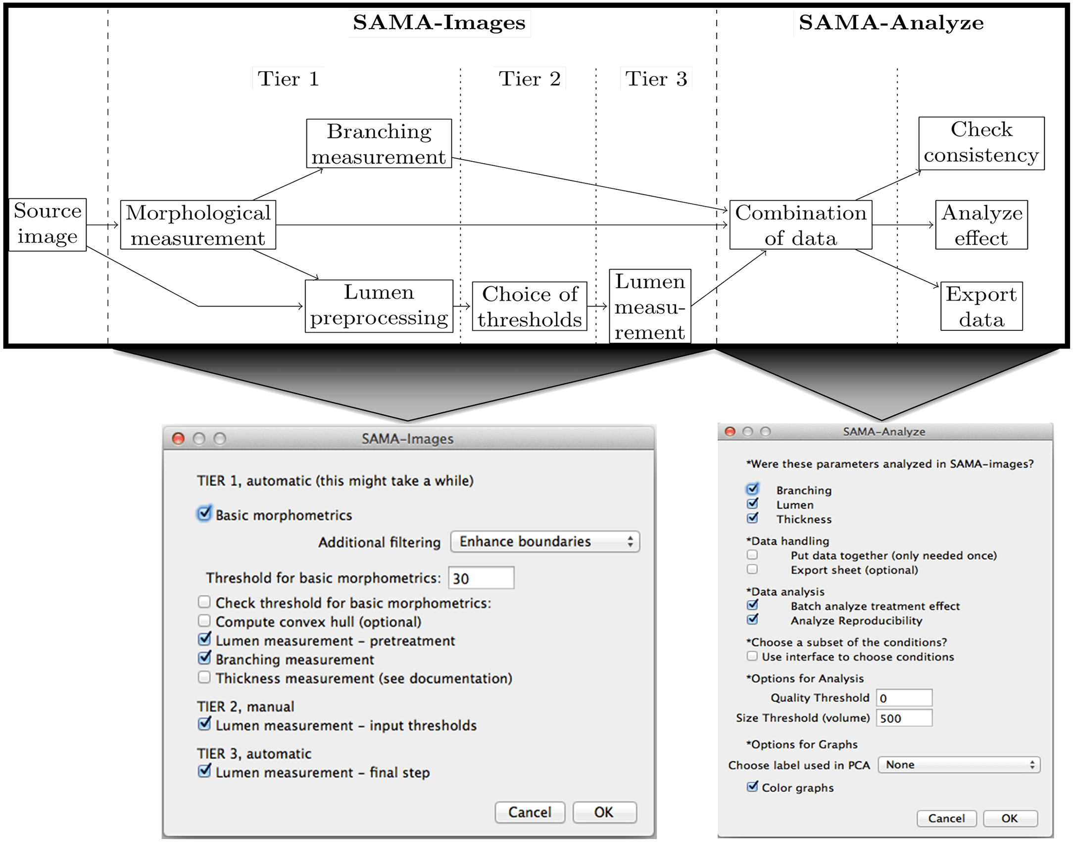
Documentation for Software for Automated Morphological Analysis : configuration and launch of the image analysis.
Documentation for Software for Automated Morphological Analysis : an overview of SAMA use and principles.
Documentation for Software for Automated Morphological Analysis : Installation of SAMA and its dependencies.
PLOS ONE


Software for Automated Morphological Analysis is a method by which epithelial structures grown in 3D cultures can be imaged, reconstructed and analyzed.
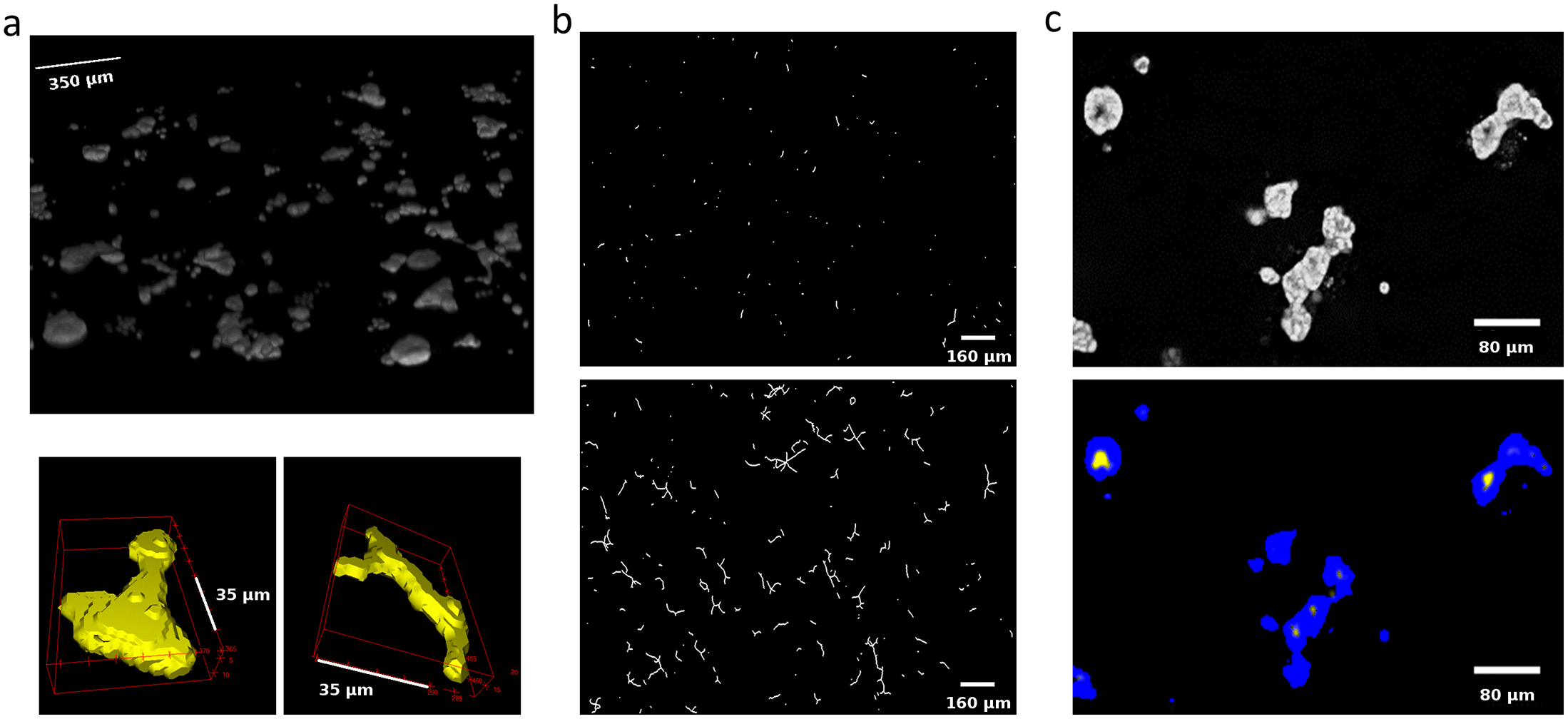
Three-dimensional (3D) culture models are critical tools for understanding tissue morphogenesis. A key requirement for their analysis is the ability to reconstruct the tissue into computational models that allow quantitative evaluation of the formed structures. Here, we present Software for Automated Morphological Analysis (SAMA), a method by which epithelial structures grown in 3D cultures can be imaged, reconstructed and analyzed with minimum human intervention. SAMA allows quantitative analysis of key features of epithelial morphogenesis such as ductal elongation, branching and lumen formation that distinguish different hormonal treatments. SAMA is a user-friendly set of customized macros operated via FIJI (http://fiji.sc/Fiji), an open-source image analysis platform in combination with a set of functions in R (http://www.r-project.org/), an open-source program for statistical analysis. SAMA enables a rapid, exhaustive and quantitative 3D analysis of the shape of a population of structures in a 3D image. SAMA is cross-platform, licensed under the GPLv3 and available at http://montevil.theobio.org/content/sama.
Keywords: Open source software, Image analysis, Ellipsoids, Morphogenesis, Computer software, Morphometry, Image processing, Branching morphogenesis
PLOS ONE
Documentation for Software for Automated Morphological Analysis, a method by which epithelial structures grown in 3D cultures can be imaged, reconstructed and analyzed.
Three-dimensional (3D) culture models are critical tools for understanding tissue morphogenesis. A key requirement for their analysis is the ability to reconstruct the tissue into computational models that allow quantitative evaluation of the formed structures. Here, we present Software for Automated Morphological Analysis (SAMA), a method by which epithelial structures grown in 3D cultures can be imaged, reconstructed and analyzed with minimum human intervention. SAMA allows quantitative analysis of key features of epithelial morphogenesis such as ductal elongation, branching and lumen formation that distinguish different hormonal treatments. SAMA is a user-friendly set of customized macros operated via FIJI (http://fiji.sc/Fiji), an open-source image analysis platform in combination with a set of functions in R (http://www.r-project.org/), an open-source program for statistical analysis. SAMA enables a rapid, exhaustive and quantitative 3D analysis of the shape of a population of structures in a 3D image. SAMA is cross-platform, licensed under the GPLv3 and available at http://montevil.theobio.org/content/sama.
Keywords: Open source software, Image analysis, Ellipsoids, Morphogenesis, Computer software, Morphometry, Image processing, Branching morphogenesis
See all tags.